Aetiology and Risk factors Epiretinal membrane has also been termed as macular pucker, epimacular membrane, preretinal macular fibrosis, surface-wrinkling retinopathy. It is a semi-translucent fibrocellular tissue proliferates on the surface of the internal limiting membrane.
Although clinical ERMs presentation are mostly idiopathic with older age, it may be present following intraocular surgery, trauma, retinal vascular diseases, retinal tear or detachment, ocular inflammatory disease, posterior vitreous detachment.
Pathophysiology It has been postulated that vitreous degenerative changes lead to the proliferation of glial cells, and the inflammatory mediators may also promote fibrocellular growth resulting to ERM formation.
Visual Acuity then and now I have no pre-existing ocular and systemic condition. I started noticing a slight distance and near blurriness in the right eye incidentally during my vacation 9 years ago. I was guessing this to be the result of cataract as the slight blurred vision was stable and no difference of colour perception in both eyes throughout my two weeks of holiday without any other sign and symptom.
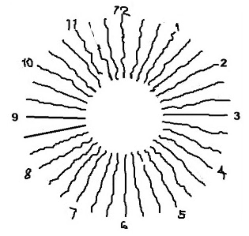
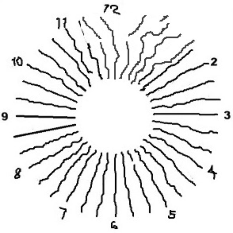
After returned home, a quick self-test found my right eye best corrected VA was at 6/12 level. When looking at the fan-chart, I shockingly saw all radiating lines appeared wavy (Fig1) and the clarity of the lines fluctuating on eye blink. My clinical instinct told me that I was having ERM.
Fig 1
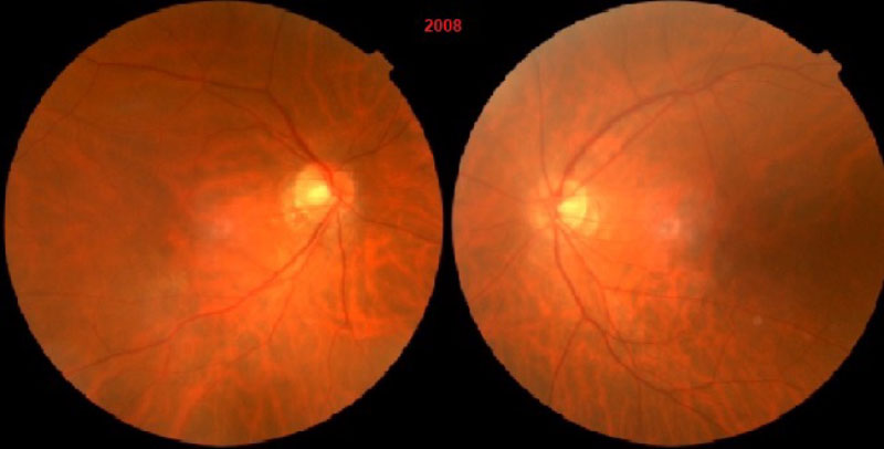
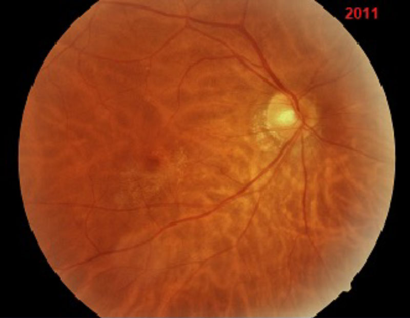
Fig 2
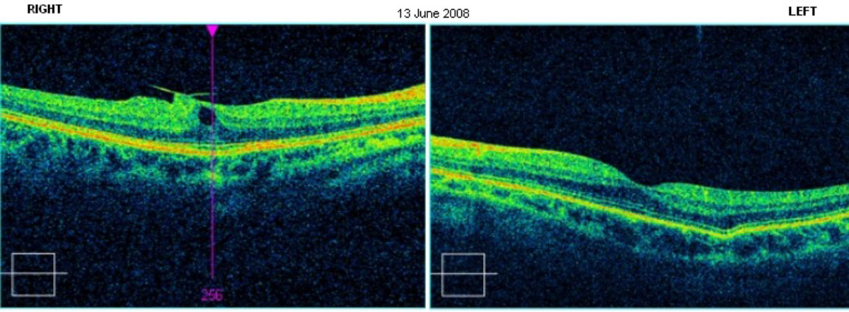
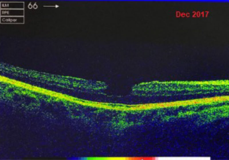
Although fundus pictures (Fig 2) revealed no obvious indication of ERM, a thorough dynamic retinal examination by a Vitreo-Retinal ophthalmologist and the Optical Coherence Tomography scan image (Fig 3) confirmed I was having ERM without any other problem, and advised to be vigilant for any deterioration of vision.
Fig 3
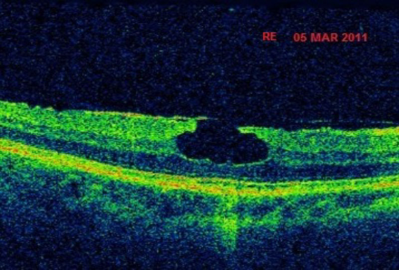
The OCT scans showing the ERM tugging on macular Three years later, my right VA deteriorated slightly to 6/15, the OCT scan image (Fig 4) showed an enlarged macular, the lamellar hole, with photoreceptors remain, more wrinkling and a coarser membrane overlaying the macular. The fundus picture showed obvious macular puckers (Fig 5). Section of the fan-chart’s radial lines (Fig 6) appeared wavier than the others, some lines appeared fractured or broken and changes in position on blinking.
Fig 4
Fig 5
Fig 6
Watchful waiting
Surgical intervention is advised when visual functions affecting normal daily activities.
The only treatment option for ERM is delicate surgical peeling of the membrane, which also involving vitrectomy, possible pneumatic retinopexy and head posturing, and cataract surgery.
Fortunately, my right eye VA improving slowly after the peak of my worst VA in my life, to the present 6/6 level, indicating to a spontaneous recovery from the ERM. (Fig 7 & 8)
Fig 7
Fig 8
Note: In the absence of novel ophthalmic OCT scanner and a poor fundus view through the undilated pupils, Fan-Chart may be the simplest diagnostic test for ERM.